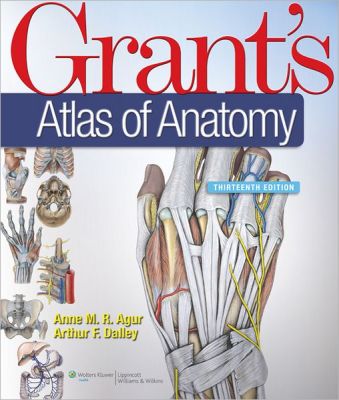
Grant's Atlas of Anatomy, 13th Edition, International edition
by Anne M.R. Agur-
Softcover
KWD17.050

A cornerstone of gross anatomy since 1943, Grant's Atlas of Anatomy reaches students worldwide with its realistic dissection illustrations, detailed surface anatomy photos, clinical images and comments, and quick-reference muscle tables. Renowned for its accuracy, pedagogy, and clinical relevance, this classic atlas boasts significant enhancements, including updated artwork, new conceptual diagrams, and vibrantly re-colored illustrations. Clinical material is clearly highlighted in blue text for easy identification.
The book contains predominantly color illustrations, with some black-and-white illustrations.
For over 50 years, this publication has been the lab partner of students who appreciate fine artwork, realistic presentation, and concise observations. It features regional organization presented in a sequence based on how the reader would perform an actual laboratory dissection. This tenth edition includes increased sizing of many figures, more information on neuroanatomy, and new features such as tables of muscles, and an MRI section at the end of each chapter.
Overview
A cornerstone of gross anatomy since 1943, Grant's Atlas of Anatomy reaches students worldwide with its realistic dissection illustrations, detailed surface anatomy photos, clinical images and comments, and quick-reference muscle tables. Renowned for its accuracy, pedagogy, and clinical relevance, this classic atlas boasts significant enhancements, including updated artwork, new conceptual diagrams, and vibrantly re-colored illustrations. Clinical material is clearly highlighted in blue text for easy identification.
The book contains predominantly color illustrations, with some black-and-white illustrations.
For over 50 years, this publication has been the lab partner of students who appreciate fine artwork, realistic presentation, and concise observations. It features regional organization presented in a sequence based on how the reader would perform an actual laboratory dissection. This tenth edition includes increased sizing of many figures, more information on neuroanatomy, and new features such as tables of muscles, and an MRI section at the end of each chapter.


